.gif)

|
Imaging in Developing Countries |
|
Special Interest Group |


|
The x-ray department was definitely the most basic and primitive I have ever seen. The x-ray unit was more than 30 years old and when I arrived they were using wet developing. Having only used CR for most of my time as a radiographer I was unable to help the two x-ray technicians improve their radiographic technique as they were already working to a high standard with the limited resources available to them (despite having no formal radiography training!). The majority of the x-ray workload was surprisingly from RTAs, most commonly motorcycle accidents. The poor quality of the roads and lack of protective clothing or helmets meant horrific injuries were a daily occurrence. |
|
|
|
Several weeks before my arrival, the hospital had received an AGFA Curix automatic x-ray processor and a Phillips BV25 image intensifier from Northern Ireland. |
|
|
|
Image intensifiers are few and far between in Uganda- there are eight in the entire country with a population of 25 million! There was therefore considerable excitement concerning the use of the machine. |
|
Fortunately I had previously had plenty of experience using a similar image intensifier in the UK and was able to get it up and running fairly easily. I taught two of the staff how to use it and wrote them an instruction guide and a diagram of how the buttons work. We managed to perform retrograde pyelography and a micturating cystogram under fluoroscopy control. Perhaps the greatest difficulty was trying to emphasise the ALARA principle and the importance of radiation protection as the technicians were fairly ‘trigger happy’ with the exposure button! |
|
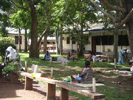
Report on Visit to Kiwoko Hospital, Uganda |
|
The staff at the hospital had also anticipated that I would be able to install the x-ray processor. This was also well out of my comfort zone however after some internet research and several long and expensive phone calls to AGFA and one of my X-ray engineer friends in the UK, I managed to get the processor working in the tiny darkroom. It was very humbling to hear the two x-ray technicians saying that they had seen such things in books but had only dreamt of using one! |
|
One of the greatest frustrations in both X-ray and Ultrasound was the lack of reliable electricity. While the hospital had it’s own generator, this was only turned on for several hours each day and the rest of the time we relied on the national grid. There would often be no electricity at all from 4pm until 9am the following morning. |
|
|
|
While my accommodation was of a high standard for this area of Uganda, it took a while to get used to using an outdoor ‘latrine’ for a toilet and sharing a bedroom with a host of cockroaches and mosquitoes! |
|
|
|
Overall, although I was faced with many challenges, I found my experience in Uganda invaluable and would recommend an elective like this to any student or qualified radiographer/sonographer. |